|
Noninvasive Cellular Imaging of the Skin
The VIVASCOPE 1500 reflectance confocal imaging system offers a non-invasive way to
image the skin in vivo from the surface to the superficial collagen layers.*
Features
- Capture images up to 8 x 8
- Macro and micro imaging
- FDA 510(k) cleared
Literature

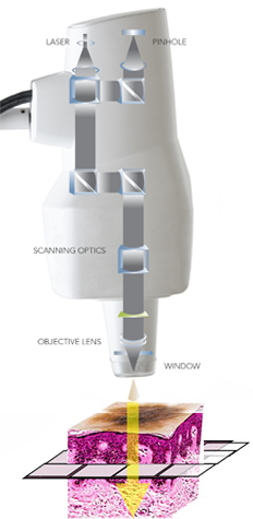 |
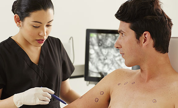
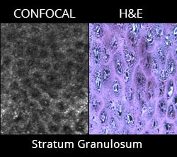
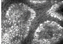
CONFOCAL IMAGING
The VIVASCOPE® system is an in vivo
confocal imaging tool that uses a lowpowered
laser to provide non-invasive,
real-time, high-resolution images of
the epidermis and the superficial
collagen layers.* The system provides
optical sectioning of unstained
epithelium and the supporting stroma
at a cellular level that may be reviewed
by a physician to assist in forming a
clinical judgment. |
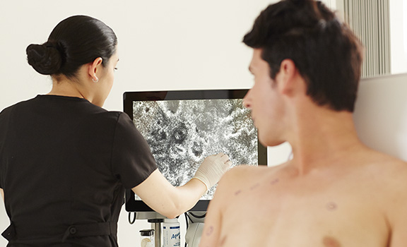
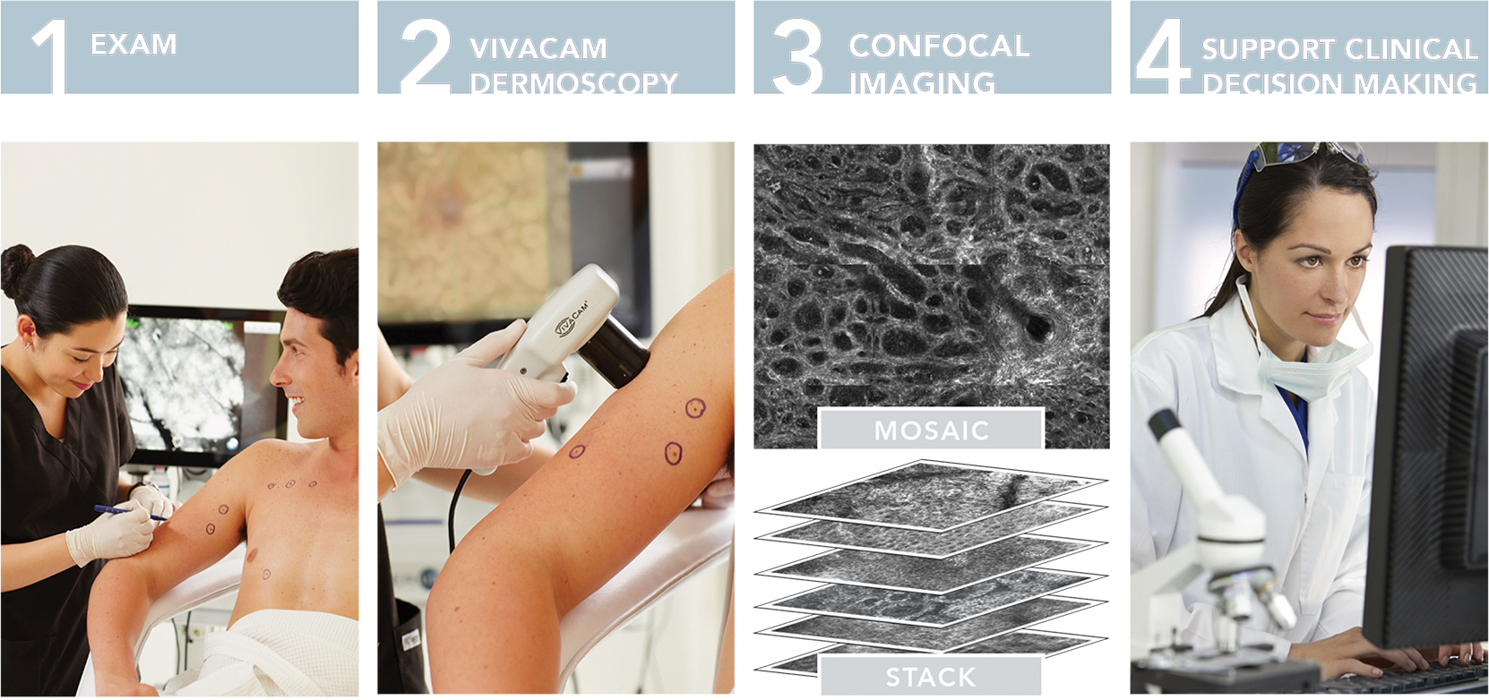
VIVACAM® captures both clinical and dermoscopic images with a high-precision optical system.
- High-resolution images
- Use to navigate within the macro image to specified corresponding confocal image
|
 |
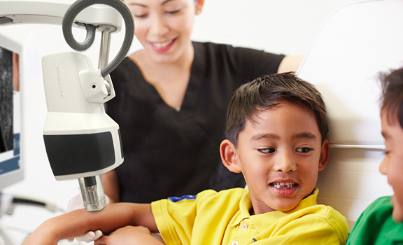
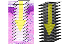
The VIVASCOPE 1500 captures single images of skin that are parallel to the surface or on the “horizontal plane”.
The image plane, or position within the skin, is changed by moving the objective lens up, down or laterally, relative to the skin surface. |
 |
| A series of horizontal images captured in “Z-depth”, or from the surface of the skin to the superficial collagen, parallel to the skin surface. The image plane is incrementally changed by moving the objective lens of the VIVASCOPE 1500, and images are captured at consecutively deeper depths within the skin. |
 |
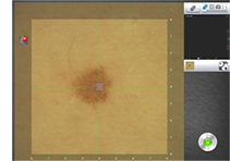
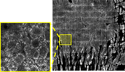
| An array of images taken on a single horizontal plane creates a mosaic. The VIVASCOPE 1500 scans the objective lens laterally over the skin while capturing a series of images and displaying them as a composite image covering an area up to 8 x 8mm. |
 |
2018 Medicare Reimbursement for RCM
The Current Procedural Terminology (CPT)* codes used for reimbursement for RCM (Reflectance Confocal Microscopy) procedure are:
- 96931 Reflectance confocal microscopy (RCM) for cellular and sub-cellular imaging of skin; image acquisition and interpretation and report, first lesion
- 96932 image acquisition only, first lesion
- 96933 interpretation and report only, first lesion
- 96934 image acquisition and interpretation and report, each additional lesion
- 96935 image acquisition only, each additional lesion
- 96936 interpretation and report only, each additional lesion
For a presentation on reimbursement by Daniel Mark Siegel MD, MS, FAAD, Clinical Professor of Dermatology, SUNY Downstate, click here.
*CPT codes and descriptions only are copyright 2017 American Medical Association (AMA). All rights reserved. The AMA assumes no liability for data contained or not contained herein.
The coding, coverage, and payment information contained herein is gathered from various resources and is subject to change without notice. Caliber Imaging & Diagnostics, Inc. cannot guarantee success in obtaining third-party insurance payments. Third-party payment for medical products and services is affected by numerous factors. It is always the provider’s responsibility to determine and submit appropriate codes, charges, and modifiers for services that are rendered. Providers should contact their third-party payers for specific information on their coding, coverage, and payment policies.
|
HIGHLIGHTS
- Non-invasive image capture
- Real-time cellular-level images of tissue
- VIVACAM Images
- CE Mark and FDA 510(K) cleared
|
|
Have questions about reflectance confocal microscopy (RCM) reimbursement?
Click here to watch a presentation given by Daniel Mark Siegel MD, MS, CPCD, Clinical Professor of Dermatology, SUNY Downstate. |
|