

VivaCam® captures both clinical and dermoscopic images with a high-precision optical system that works to avoid color, brightness variations, and geometric distortions. Photo quality overview shots can be used to document pre-operative and post-operative treatment statuses.
THE INTELLIGENT CAMERA
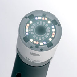
Unique integrated microprocessors adjust the exposure intensity and color mixture automatically to provide uniform, standardized image quality in all magnifications. Highly detailed, true-to-life, best quality color images are produced within seconds. Microprocessor-controlled mixture of light allows for the most precise diagnostic microscopic examination of skin lesions.
The single camera system allows for smooth navigation between overview and dermoscopic images. This time-saving concept allows users to focus their concentration primarily on the patient. Using the macro function allows for large skin areas to be documented without the inconvenience of switching cameras or camera lens.
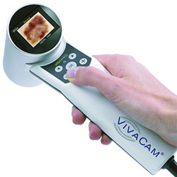
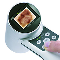
The skin area being examined is displayed on both the camera’s built-in OLED display and the VivaScan software. Clinical and dermoscopic images are stored for review and annotation within the VivaScan® software. With VivaCam®, minimal changes in pigmented lesions can be reliably visualized and documented over time.
CONFOCAL & DERMOSCOPY
The VivaCam produces an image with a 10 mm field of view. Linking the VivaCam with the VivaScope® System allows clinicians and researchers to navigate within the macro image to specify the subsequent confocal imaging with the VivaScope.